
Cachan & Paris 13
Technical platform: our diagnostic equipment
Two practices, one principle: measure before deciding.
A technical platform is not a showroom of hardware: it is what turns a visual complaint into a precise diagnosis, and then into a reasoned treatment decision. The Cachan and Paris 13 practices are equipped with complementary instruments: corneal imaging, retinal tomography, biometry, visual field testing, ultra-widefield retinal imaging and treatment lasers. Cornea, cataract, glaucoma and retina are all managed at both sites; only some retinal imaging equipment is concentrated at Paris 13. This page explains what each test measures, when it is requested, and what it contributes to your pre-operative assessment or to your follow-up.
UNDERSTANDING
Why the equipment matters as much as the surgery itself
In refractive surgery, as in cataract surgery, the quality of the outcome is largely decided before the operating theatre. The procedure itself lasts a few minutes; what makes it suited to your eye are the measurements that preceded it.
Three concrete examples. A corneal topography showing early irregularity may lead us to rule out LASIK in favour of PRK, or to postpone surgery altogether. Imprecise biometry translates into an error in lens power, and therefore into residual spectacle dependence after cataract surgery. A macular OCT that reveals an epiretinal membrane completely changes the visual prognosis given to the patient, and with it the discussion about which lens to choose.
In all three situations, the instrument did not replace clinical reasoning: it provided a measured, reproducible figure that allows a decision to be discussed with you rather than announced to you. This is also why certain tests are repeated over time: how a measurement changes, rather than its value on a single day, carries the greater diagnostic weight.
CACHAN PRACTICE
The Cachan equipment
The Cachan practice (consultations on Mondays, Wednesdays and Fridays) handles general ophthalmology consultations, pre-surgical work-up for refractive and cataract surgery, glaucoma monitoring and retinal assessment. Seven instruments are installed there.
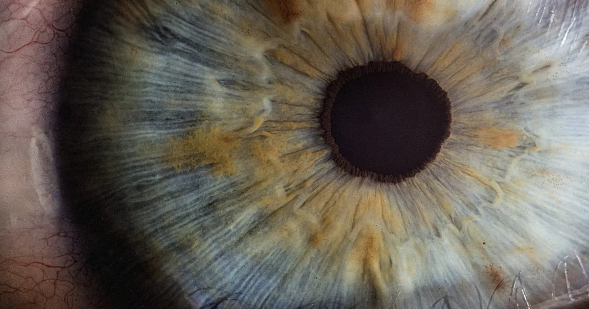
Sirius+ topographer (CSO) — the map of your cornea
The Sirius+ combines a Placido disc with a rotating Scheimpflug camera. It produces a map of both the anterior and the posterior surface of the cornea, together with a thickness map (pachymetry) across the whole surface, where an ultrasound pachymeter gives only a single measurement point.
This is the key test for detecting keratoconus and its forme fruste or subclinical variants, which rule out LASIK. Analysing the posterior surface together with the thickness distribution improves the detection of early forms compared with anterior curvature alone (PMID 32832241). Topography is also used to measure corneal astigmatism before a toric lens is implanted, and it provides corneal aberrometry: the measurement of higher-order optical defects, which cannot be corrected with conventional spectacles and which explain some complaints of halos or degraded night vision. This test is described in detail on the corneal topography page.
Cirrus 6000 OCT (Zeiss) — a cross-section of the retina
Optical coherence tomography produces a cross-section of the retina at a resolution of around 5 µm, without contact and without injection. The Cirrus 6000 acquires 100,000 scans per second, which reduces artefacts caused by eye movement.
There are two main uses. At the macula, it detects oedema, an epiretinal membrane, a macular hole or early AMD — all conditions that change the prognosis of cataract surgery. At the optic nerve, it measures the thickness of the nerve fibre layer and of the macular ganglion cells, parameters used for diagnosing and monitoring glaucoma, with diagnostic performance comparable between macular analysis and peripapillary nerve fibre analysis (PMID 26891880).
IOL Master 700 (Zeiss) — calculating the cataract lens
Biometry measures the axial length of the eye, corneal curvature, anterior chamber depth and lens thickness. These values feed the calculation formulas that determine the power of the intraocular lens implanted during cataract surgery.
The IOL Master 700 uses swept-source OCT technology, which penetrates dense cataracts better than previous-generation optical biometry, and displays a full cross-section of the eye that can reveal eccentric fixation. Studies comparing swept-source biometers find very similar power predictions between current devices (PMID 38643892). The precision of this measurement is decisive when a premium multifocal or toric lens is being considered.
Humphrey Field Analyzer 3 (Zeiss) — peripheral vision
Automated perimetry tests retinal sensitivity at around fifty points, one eye at a time. It documents deficits that the patient cannot yet perceive — glaucoma progresses for a long time without symptoms — and makes it possible to follow their evolution from one year to the next.
Visual field testing is also requested when investigating certain neuro-ophthalmic conditions, and it is required for driving fitness in several licence categories.
Tonoref III (Nidek) — four measurements in one pass
The Tonoref III combines an autorefractor, a keratometer, a non-contact tonometer and a pachymeter. In a few seconds it provides a baseline objective refraction, corneal curvature, intraocular pressure and central corneal thickness.
That last point is far from incidental: a thin cornea leads to underestimating the measured pressure, a thick cornea to overestimating it. Pachymetry therefore puts the pressure reading back into context, an essential step before discussing ocular hypertension or glaucoma.
Tango laser (Ellex) — three treatments, one platform
The Tango combines a YAG laser and an SLT laser. It allows three procedures to be carried out in the consulting room, without hospital admission: YAG capsulotomy for secondary cataract, peripheral iridotomy to prevent angle-closure glaucoma, and selective laser trabeculoplasty (SLT) to lower intraocular pressure in open-angle glaucoma.
Nidek slit lamp — the reference examination
No imaging replaces biomicroscopy. The slit lamp gives a three-dimensional view of the eyelids, the conjunctiva, the cornea layer by layer, the anterior chamber, the iris and the crystalline lens — it is what allows the type and density of a cataract to be graded. Combined with an examination lens, it also gives access to the fundus and to the iridocorneal angle.
The slit lamp is also where the tear film is assessed, and this has a direct practical consequence: untreated dry eye distorts pre-operative measurements and increases discomfort after corneal surgery. It is therefore managed before the procedure, not after.
PARIS 13 PRACTICE
The Paris 13 equipment: widefield imaging and laser
The retina is assessed at both sites. The Paris 13 practice, which also hosts the consultations dedicated to screening in patients with diabetes, additionally brings together widefield retinal imaging and the photocoagulation laser: this is where fundus assessments and treatments are directed.
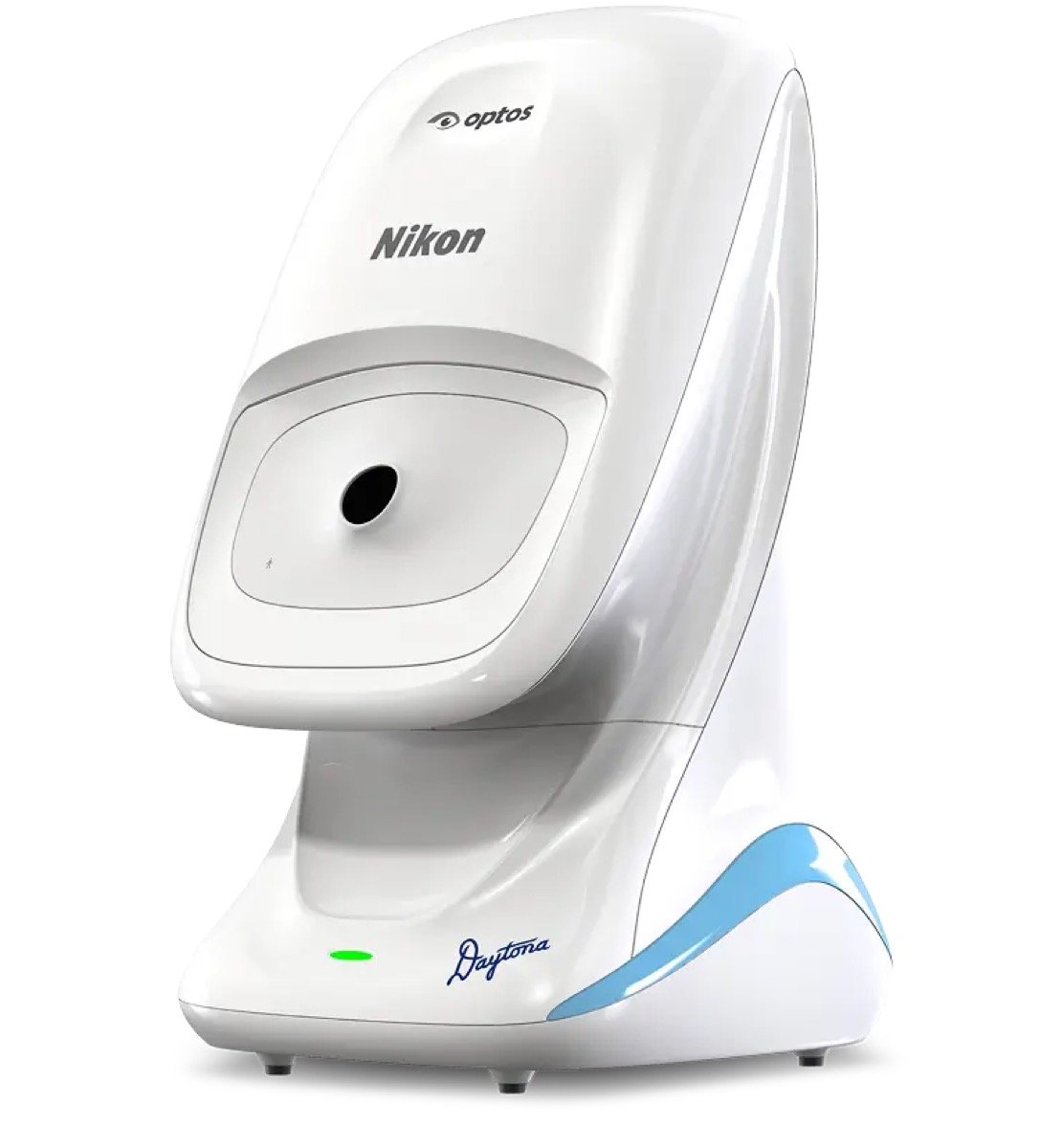
Optos Daytona — around 200° of retina in one image
A conventional retinal camera photographs 45 to 50° of the fundus, that is, the posterior pole. The Optos Daytona uses confocal scanning laser imaging and an ellipsoidal mirror to capture around 200°, in the region of 80% of the retinal surface, in a capture lasting less than a second — usually without pupil dilation.
The benefit is twofold. First, the retinal periphery becomes visible in a single image: this is where tears, lattice degeneration and part of the lesions of diabetic retinopathy are found. Studies comparing ultra-widefield imaging with ETDRS 7-field stereoscopic photography, long regarded as the reference standard, report good agreement for grading diabetic retinopathy (PMID 22912430).
Second, comfort: not needing dilation in most cases avoids several hours of blurred vision and allows patients to drive home. The examination also provides an archived baseline image against which later checks can be compared point by point.
Toward Pi Yalkaid OCT — angiography without injection
The Yalkaid is a swept-source OCT running at 100 kHz, with an OCT angiography (OCT-A) module. This second modality reconstructs the retinal and choroidal circulation by analysing the flow signal, without any fluorescein injection, and therefore without the allergic risk and the nausea associated with conventional angiography.
In practice, OCT-A shows areas of capillary non-perfusion and early new vessels in diabetic retinopathy and AMD. The longer wavelength of swept-source technology penetrates hazy media and the choroid better than spectral-domain OCT. Recent developments are extending the field of OCT angiography towards the retinal periphery (PMID 36383595). Fluorescein angiography nevertheless retains its own indications, in particular for studying leakage.
Quantel laser platform — SLT, YAG and the Vitra module
The Paris 13 practice has a Quantel Medical laser platform that brings together two anterior segment procedures performed in the consulting room: YAG capsulotomy, when clouding of the capsule causes vision to fall again after cataract surgery, and selective laser trabeculoplasty (SLT), which lowers intraocular pressure in open-angle glaucoma. The YAG laser also allows peripheral iridotomy to prevent angle-closure glaucoma.
A Vitra module completes this platform for retinal photocoagulation. It treats tears and peripheral lesions with a laser barrier, and covers the indications of diabetic retinopathy when the disease warrants it. Treatment is guided by imaging performed the same day: widefield retinal imaging locates the areas to be treated, and OCT confirms the absence of associated macular oedema.
Tonometry and digital slit lamp
The site also has a Tonoref III and a slit lamp with digital imaging, which makes it possible to photograph an anterior segment lesion and to follow its evolution objectively from one consultation to the next.
IN PRACTICE
Which test for which situation?
Tests are not ordered as a routine batch: each answers a specific clinical question. Here are the most frequent combinations.
You are considering refractive surgery
Sirius+ topography, VX40 aberrometry, pachymetry, refraction under cycloplegia, fundus examination. The aim is twofold: to check that the cornea can tolerate the treatment, and to choose between LASIK, PRK and SMILE.
A cataract has been diagnosed
IOL Master 700 biometry, topography if a toric lens is being considered, macular OCT to check the state of the macula before giving a visual prognosis.
You have diabetes
Optos ultra-widefield retinal imaging, macular OCT to look for oedema, OCT angiography if vascular involvement is suspected.
Raised eye pressure has been detected
Pachymetry to interpret the pressure reading, OCT of the optic nerve and ganglion cells, Humphrey visual field. These three tests are then repeated to judge whether anything is progressing.
You see floaters or flashes of light
Dilated fundus examination and widefield retinal imaging, looking for a peripheral retinal tear. This situation warrants a prompt consultation.
Your central vision is distorted
Macular OCT as the first-line test: it distinguishes an epiretinal membrane, macular oedema and wet AMD, which have neither the same prognosis nor the same treatment.
What these instruments do not do
It would be misleading to suggest that a technical platform is enough to make a decision. Three limits deserve to be stated.
A measurement is not a diagnosis. Corneal thinning on its own does not mean keratoconus; a pressure of 22 mmHg does not mean glaucoma. It is the consistency between several tests, together with age, family history and change over time, that builds a diagnosis.
These instruments produce false positives. Analysis software compares your values with a normative database. High myopia, an atypical optic disc or simple dry eye are enough to flag an abnormality that is not one. Interpretation remains a medical task.
Recent equipment does not guarantee an outcome. Refractive surgery and cataract surgery carry risks, low but real, which are not removed by the quality of the pre-operative measurements. These risks are explained to you in consultation, before any decision is made.
FOLLOW-UP
Your images follow you from one consultation to the next
Every capture is archived and attached to your file. This has an important practical consequence: at a follow-up visit, the instrument does not simply produce a new image, it overlays it on the previous one. The trend analysis software of the OCT and of the visual field can then calculate a rate of progression, in microns per year or decibels per year.
It is this slope, far more than a single reading, that prompts a change of treatment in glaucoma. A patient whose nerve fibres remain stable over five years and a patient who steadily loses thickness are not managed in the same way, even at identical pressures.
In concrete terms, this also means that changing instrument means losing comparability: OCT measurements are not directly transferable from one manufacturer to another. That is one of the reasons why it is preferable to continue follow-up on the same equipment, and why we ask you to bring your previous test results: even when they cannot be overlaid, they provide a numerical reference point.
Finally, the data needed in theatre — biometry, topography, astigmatism axis — is sent to the facility where the procedure takes place, so that the lens calculation and the laser settings rest on your own measurements rather than on figures re-entered by hand.
Frequently asked questions about these tests
Do my pupils need to be dilated for Optos retinal imaging?
In most cases, no: the Optos Daytona captures its image through an undilated pupil. Dilation is still requested when the examination is looking for a very peripheral lesion, when the pupil is narrow, or when an additional contact lens examination is required.
Are these tests painful?
Topography, OCT, retinal imaging, biometry and visual field testing are all non-contact examinations: no instrument touches the eye. Non-contact tonometry sends a brief puff of air, felt as surprising rather than unpleasant. Examinations performed with a contact lens are preceded by anaesthetic eye drops.
How long does a full assessment take?
A pre-operative refractive surgery assessment usually takes one to two hours on site, including dilation and waiting time. A diabetic screening assessment is shorter. The full sequence is described on the pre-operative assessment page.
Are these tests reimbursed?
Tests carried out to diagnose or monitor a medical condition (diabetes, glaucoma, AMD, cataract) fall within the French statutory health insurance schedule. By contrast, the assessment carried out for refractive surgery performed for non-therapeutic reasons is not covered; the arrangements are set out on the pricing page.
Can I drive after the consultation?
If no dilation has been performed, yes. After dilation, near vision remains blurred and sensitivity to light is increased for about four to six hours: it is preferable to arrange another means of transport and to bring sunglasses. Let us know beforehand if you absolutely must drive.
Should I bring my previous test results?
Yes, it is useful. In ophthalmic imaging, comparison over time is often more valuable than a single measurement: an OCT or a visual field carried out elsewhere two years earlier makes it possible to judge stability or progression. Please also bring your spectacle prescriptions and the list of your medications.
Is every instrument available at both sites?
No, and that is deliberate. Both practices cover cornea, cataract, glaucoma and retina. Some equipment is grouped together, however: the detailed pre-surgical assessment (topography, biometry, visual field) is carried out in Cachan, while widefield retinal imaging and the photocoagulation laser are located in Paris 13. You will be told which site to attend according to the examination required. Addresses and directions are given on the practices page.
Scientific sources
- Kernt M, Pinter F, Hadi I, Ulbig M, et al. Assessment of diabetic retinopathy using nonmydriatic ultra-widefield scanning laser ophthalmoscopy (Optomap) compared with ETDRS 7-field stereo photography. Diabetes Care. 2012;35(12):2459-2463. PMID 22912430
- Niederleithner M, de Sisternes L, Stino H, et al. Ultra-Widefield OCT Angiography. IEEE Trans Med Imaging. 2023;42(4):1009-1020. PMID 36383595
- Sorkin N, Zadok R, Savoldelli A, et al. Comparison of Intraocular Lens Power Prediction Accuracy Between 2 Swept-Source Optical Coherence Tomography Biometry Devices. Am J Ophthalmol. 2024;265:156-164. PMID 38643892
- Consejo A, Solarski J, Karnowski K, et al. Keratoconus Detection Based on a Single Scheimpflug Image. Transl Vis Sci Technol. 2020;9(7):36. PMID 32832241
- Oddone F, Lucenteforte E, Michelessi M, et al. Macular versus Retinal Nerve Fiber Layer Parameters for Diagnosing Manifest Glaucoma: A Systematic Review of Diagnostic Accuracy Studies. Ophthalmology. 2016;123(5):939-949. PMID 26891880
Further reading
- Corneal topography: what is this test for?
- The pre-operative refractive surgery assessment, step by step
- The Cachan and Paris 13 practices: addresses, directions and opening times
Cachan and Paris 13 practices · Tel. 01 45 47 08 11
Disclaimer
This page is for information only. A personalised ophthalmic opinion remains essential for any treatment decision.
The description of the equipment given here is neither a prescription nor a guarantee of outcome. The tests performed are tailored to each clinical situation and their interpretation is a matter for medical examination. The indications, the alternatives and the risks of any procedure are explained to you in consultation, before any decision is made. For any question concerning your vision, please consult an ophthalmologist.
The brand names and product photographs mentioned belong to their respective manufacturers (Zeiss, CSO, Nidek, Ellex, Optos, TowardPi Medical, Quantel Medical) and are cited descriptively to identify the equipment in use. Reference sources: the French National Authority for Health (HAS), the French Society of Ophthalmology, and the PubMed-indexed publications listed above.