
Refractive surgery: Trans-PRK & PRK
Trans-PRK & PRK in Paris and Cachan: Surface Laser Refractive Surgery
Trans-PRK (transepithelial PRK, 100% laser, no-touch) and conventional PRK are the reference surface laser techniques for thin corneas. Results identical to LASIK, with no corneal flap created.
Hesitating between the two techniques? LASIK or PRK: how to choose — our comparison covers cornea, recovery, dry eye, sport and price.
UNDERSTANDING PRK
What is PRK? The principle of photorefractive keratectomy
PRK (PhotoRefractive Keratectomy), also called surface laser, is historically the first refractive surgery technique using the excimer laser. Developed in the late 1980s, it remains a reference technique today, particularly indicated when LASIK is contraindicated.
Unlike LASIK, PRK does not require the creation of a corneal flap. The surgeon removes the epithelium (the surface cell layer of the cornea, roughly 50 µm thick) either mechanically or using a diluted alcohol solution. The excimer laser is then applied directly to the exposed corneal stroma, with the same personalized ablation profile as for LASIK. The absence of a flap is both PRK’s main advantage (corneal strength preserved) and its drawback (slower recovery, since the epithelium has to regenerate over 3 to 5 days).
At the Clinique Laser Victor Hugo (Paris 16th), Dr Tourabaly uses the Schwind Amaris 750S excimer laser for PRK and Trans-PRK. This latest-generation laser offers an optimized aspheric ablation profile, eye-tracking at 1,050 Hz and aberrometry-guided treatment to maximize postoperative visual quality. The Cachan practice handles preoperative consultations and postoperative follow-up.
PRK or LASIK: what are the differences?
PRK and LASIK use the same excimer laser and produce identical refractive results in the long term. The fundamental difference concerns access to the corneal stroma:
| Criterion | PRK | LASIK |
|---|---|---|
| Corneal flap | No: cornea intact | Yes (110 µm) |
| Visual recovery | 3 to 5 days | 24-48h |
| Postoperative pain | Moderate (3-4 days) | Very mild |
| Thin corneas | Ideal: saves 110 µm | Contraindicated |
| Corneal strength | Optimal (no flap) | Weakened by the flap |
| Contact sports | No restriction | Caution (risk of flap dislocation) |
| Final result | Identical: same laser, same precision | |
In short: if your cornea is thick enough and you want a fast recovery, LASIK is preferred. If your cornea is thin, if you play contact sports or if you work in an occupation with a risk of eye trauma (law enforcement, military, athletes), PRK is the safest choice. You can also look at the SMILE technique, which combines some of the advantages of both approaches.
Conventional PRK vs Trans-PRK: the 100% laser variant
Trans-PRK (or transepithelial PRK) is an evolution of conventional PRK that I perform in the majority of my surgical practice. The difference lies in a single step: the removal of the surface layer of the cornea, the epithelium.
In conventional PRK, the epithelium is removed manually by the surgeon: using a spatula or a diluted alcohol solution. In Trans-PRK, this removal is carried out directly by the excimer laser, in continuity with the refractive correction. The procedure thus becomes entirely no-touch: no mechanical contact with the eye, no application of alcohol.
Observed clinical advantages
- Less mechanical trauma: the cornea undergoes no direct instrumental contact.
- Improved postoperative comfort in some patients (day 0 to day 3 pain perceived as less intense, although the literature remains debated).
- No diluted alcohol on the cornea, which may be preferable for very sensitive patients.
- 100% laser protocol: optimal reproducibility from one procedure to the next.
The limits to be aware of (fair and honest information)
Recent clinical studies do not demonstrate a superior long-term benefit compared with conventional PRK in terms of final visual acuity. The choice between conventional PRK and Trans-PRK therefore depends mainly on the preferred surgical strategy and the patient’s corneal profile.
In practice, I perform the majority of procedures as Trans-PRK thanks to the Schwind Amaris 750S laser, which makes this protocol possible under optimal conditions.
Mitomycin C 0.02%: preventing corneal haze
One of the risks specific to surface surgery (PRK and Trans-PRK) is corneal haze: a fine opacity of the anterior stroma that can occur in the weeks or months following the procedure. This phenomenon is linked to healing and can, in the most pronounced cases, impair visual quality.
To prevent this risk, I systematically apply to all my patients a mitomycin C solution at a concentration of 0.02% to the corneal surface, immediately after laser photoablation. The application lasts 10 to 20 seconds depending on the ablation profile, then the cornea is copiously rinsed with balanced salt solution (BSS). This anti-haze protocol is applied regardless of the degree of myopia or astigmatism corrected.
What the scientific studies say
A meta-analysis published in 2021 evaluated visual outcomes and haze formation after intraoperative application of mitomycin C during PRK. Including 11 randomized controlled trials covering 3,536 eyes (2,232 with MMC, 1,304 controls), it concludes that there is a significant reduction in early and late haze, with better preservation of visual acuity in the treated group.
The 0.02% concentration is today the international standard for moderate to high myopia, as noted by EyeWiki and publications from the ASCRS (American Society of Cataract and Refractive Surgery).
Schwind Amaris 750S laser: 6D precision and topo-guidance
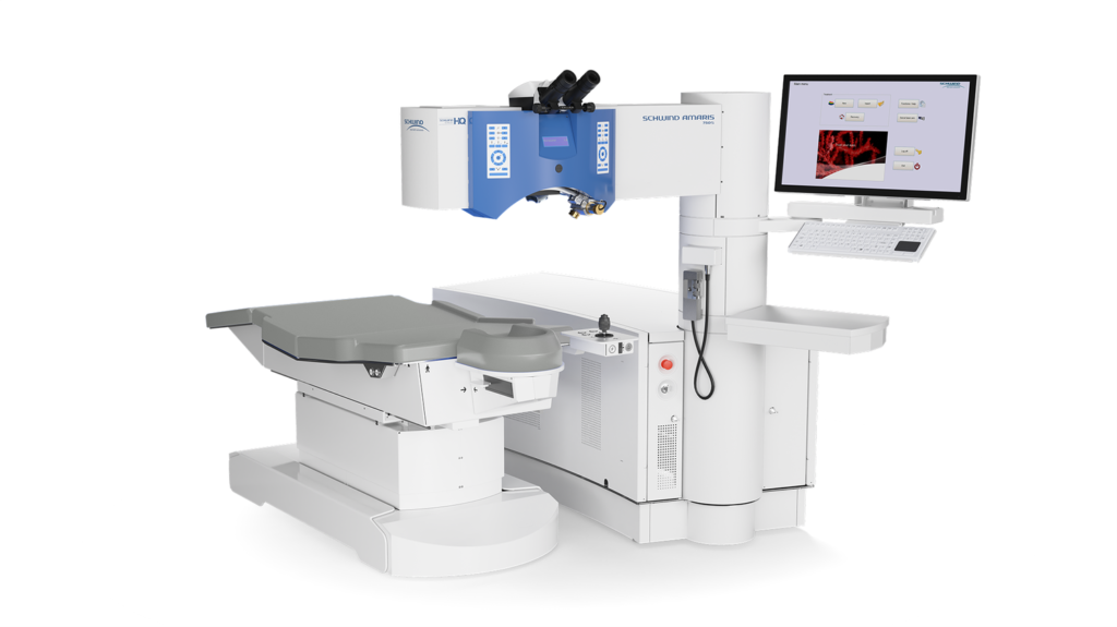
I perform my PRK and Trans-PRK procedures at the Clinique Laser Victor Hugo (Paris 16), equipped with the Schwind Amaris 750S excimer laser: one of the most high-performing platforms currently available for refractive surgery.
Speed and gentleness
The Schwind Amaris 750S delivers laser pulses at a frequency of 750 Hz, which allows it to correct one diopter in about 1.5 seconds. For a myopia of -4 diopters, the actual stromal treatment time is under 10 seconds per eye. This speed reduces corneal exposure and improves intraoperative comfort.
Six-dimensional eye-tracking
During the ablation, the eye is never completely still. The Schwind Amaris 750S tracks its position 1,050 times per second, with a latency under 3 milliseconds, across 6 dimensions:
- The horizontal and vertical movements (X and Y axes)
- The rolling movements of the eye (tilts)
- Cyclotorsion: rotation of the eye around the optical axis, both static (difference between sitting and lying positions) and dynamic (movement during the procedure). This is Advanced Cyclotorsion Control.
- The depth (Z axis), compensated by z-tracking.
This multidimensional compensation is particularly important for astigmatism corrections, where the exact orientation of the cylindrical axis determines the quality of the final result.
Personalized topo-guided ablation
Beyond standard correction, the Schwind Amaris 750S enables a topo-guided ablation: the reshaping profile is calculated from the patient’s actual corneal topography, rather than from an averaged statistical model. This makes it possible to treat both the refractive error and the patient’s individual corneal irregularities (higher-order optical aberrations) at the same time, for optimal visual quality in challenging conditions (night driving, low light).
Complementary technologies
- SmartPulse Technology: optimizes pulse distribution for a smoother post-ablation corneal surface, improving early visual recovery.
- Automatic Fluence Level Adjustment: 80% of the ablation performed at high fluence (speed), the final 20% at low fluence (precision and gentle finishing).
- Intelligent Thermal Effect Control: continuous monitoring of corneal temperature, kept below a 5°C rise to preserve the tissue.
- Particle aspiration: integrated system that maintains stable microclimatic conditions during the ablation.
Ideal candidates and contraindications for PRK
PRK is indicated to correct myopia (up to -6 diopters), hyperopia (up to +3 diopters) and astigmatism (up to 4 diopters). It is particularly recommended in the following situations:
- Thin cornea (pachymetry < 500 µm): PRK saves the 110 µm of the LASIK flap, allowing treatment of patients who are not eligible for LASIK
- Corneal surface abnormality: epithelial dystrophy, superficial corneal scars
- Occupations with a risk of eye trauma: military, police, firefighters, high-level athletes (boxing, rugby, martial arts)
- Forme fruste keratoconus: in certain borderline cases, combined with corneal cross-linking (CXL+PRK)
- Enhancement after LASIK: when an enhancement is needed and a new flap is undesirable
The contraindications are similar to those for LASIK: established keratoconus, progressive ocular disease, pregnancy, uncontrolled autoimmune disease. For myopia greater than -6 D, Dr Tourabaly will instead direct you toward LASIK (if the cornea allows it) or a phakic implant.
Step-by-step PRK procedure
Trans-PRK (no-touch), step by step
A 100 % laser surgery, with no cutting and no instrumental contact on the cornea.
-
1
Transepithelial photoablation
The excimer laser itself removes the thin surface layer (epithelium), with no instrument and no contact: this is the “trans-PRK” technique, 100 % laser.
-
2
Reshaping the stroma
At the same time, the laser reshapes the surface of the cornea to within a micron, according to the intended correction (for myopia: a central flattening).
-
3
Placing a bandage contact lens
A soft contact lens is placed like a dressing: it protects the surface and supports comfort during healing.
-
4
Re-epithelialization
The epithelium grows back spontaneously within a few days under the lens, which is then removed at a check-up visit.
-
5
Recovery
Vision stabilizes gradually over a few weeks. PRK is preferred when the cornea is thin or the activity is exposed to a risk of impact.
Surgical video · Dr Tourabaly’s practice
Trans-PRK: photoablation with the Schwind excimer laser
33 seconds of a Trans-PRK procedure on the Schwind Sirius platform. On the left, the eye-tracker (red circle) stays centered on the pupil at all times while the laser reshapes the cornea. On the right, the intraoperative view shows the astigmatism correction markers.
- Topical anesthesia: Anesthetic eye drops instilled a few minutes before. The eyelid speculum is put in place.
- Removal of the epithelium (30 seconds): The corneal epithelium is gently removed by mechanical brushing or by applying a 20% diluted alcohol solution for 20 seconds. The corneal stroma is exposed, ready for the laser.
- Excimer laser ablation (20-50 seconds): The Schwind Amaris 750S laser reshapes the surface of the stroma according to the personalized ablation profile. The 7D eye-tracker compensates for eye movements in real time. The treatment is guided by the data from the aberrometry collected during the assessment.
- Application of Mitomycin C: Systematic application to all patients of a 0.02% Mitomycin C solution for 10 to 20 seconds, immediately after photoablation, to prevent the occurrence of “haze” (scarring opacification of the stroma).
- Placing the bandage contact lens: A soft therapeutic lens is placed on the cornea to protect the treated area while the epithelium regenerates (3 to 5 days). It will be removed at the practice during the check-up.
The procedure takes 10 to 15 minutes for both eyes. The patient goes home with treatment by antibiotic, anti-inflammatory and lubricating eye drops.
CLINICAL RESULTS
Recovery and results after PRK
Recovery after PRK is more gradual than after LASIK, because the epithelium has to regenerate completely:
- Day +1 to Day +3: Moderate eye discomfort: burning sensation, watering, light sensitivity. Blurred vision. Oral painkillers prescribed if needed. This is the most uncomfortable phase.
- Day +3 to Day +5: The epithelium has regenerated. The therapeutic lens is removed at the practice during a scheduled check-up between 3 and 5 days after the procedure. The discomfort fades quickly after removal. Vision still fluctuating.
- Week +2: Significant improvement. Most patients can resume screen work. Vision at 6/10 to 8/10.
- Month +1: Vision stabilized for the majority of patients. 90% reach 10/10 without correction.
- Month +3 to Month +6: Final refractive result. Any haze (very rare with Mitomycin C) resorbs completely.
The final result of PRK is strictly identical to that of LASIK. The only difference is the recovery time: 3-5 days of discomfort instead of a few hours. In return, the cornea is more resistant and the risk of flap-related complications is eliminated.
Curcă 2025 (Trans-PRK, 137 eyes)
since 1987 (founding technique)
no mechanical contact and no alcohol
SURGICAL EXPERTISE
Why choose Dr Tourabaly for your PRK?
- Expertise in surface surgery: Dr Tourabaly is proficient in conventional PRK, trans-PRK (all-laser ablation with no mechanical contact) and combined PRK+CXL
- Latest-generation laser: Schwind Amaris 750S at the Clinique Laser Victor Hugo, with an aspheric ablation profile that minimizes postoperative aberrations
- Ultra-comprehensive assessment: The PRK vs LASIK decision is made on the basis of objective data: Pentacam topography, pachymetry, aberrometry
- Close follow-up: Check-ups at Day +1, Day +3 to Day +5 (removal of the therapeutic lens at the practice), then Week +2, Month +1, Month +3 and Month +6
- More than 1,000 Google reviews: 4.9/5: see patient testimonials
Where do the procedures take place?
Refractive surgeries are performed at:
• Clinique Laser Victor Hugo: 27 bis avenue Victor Hugo, 75116 Paris (operation-yeux-laser.com)
The preoperative assessment takes place at the Cachan practice (94).
FAIR AND HONEST INFORMATION
Possible complications and how they are managed
Like any surgical procedure, PRK carries risks that it is essential to understand before committing. I systematically present these risks during the consultation, as a duty of fair and honest information. The vast majority remain rare or transient. To learn more, see our summary of the safety data on refractive surgery.
Postoperative pain (Day 0 to Day 3)
The healing of the epithelium (48 to 72 hours) is accompanied by discomfort in the form of stinging, watering and sensitivity to light. These symptoms are expected and temporary. They are eased by a bandage contact lens worn until Day +4, and by a local painkilling and anti-inflammatory treatment prescribed as standard.
Corneal haze
A fine opacity of the anterior stroma, which can occur in the weeks following the procedure. As detailed above, the systematic application of mitomycin C 0.02% during the procedure significantly reduces this risk. Visually significant haze remains exceptional today.
Under-correction or over-correction
The individual biological response to photoablation can vary. In rare cases, the final correction deviates from the target, which may justify an enhancement (after refractive stabilization, generally 6 to 12 months post-op). The precision of the Schwind Amaris 750S and the thorough preoperative assessment limit this risk.
Delayed epithelial healing
The corneal epithelium usually reconstitutes itself in 3 to 5 days. In some patients (pre-existing dry eye, ocular surface disease), this time may be prolonged. Close follow-up allows the treatment to be adjusted where necessary.
Transient dry eye
The very first postoperative weeks may be marked by dry eye, linked to the transient change in corneal innervation. This symptom is generally less pronounced than after LASIK, and resolves within a few weeks to a few months with artificial tears. It is precisely because PRK does not create a corneal flap — and therefore better preserves the deep innervation of the cornea — that it is often preferred in patients with pre-existing dry eye, despite a slightly more sensitive surface-healing phase in the very first days.
Nighttime visual perceptions
Halos or glare in night vision may be perceived in the first weeks. They are overall less frequent with PRK than with LASIK (no corneal flap) and subside in the vast majority of cases as healing stabilizes.
Infection (keratitis)
An exceptional complication (estimated incidence under 1/5,000). The bandage contact lens, postoperative antibiotic therapy and strict adherence to hygiene instructions minimize this risk. Any unusual symptom (intense pain, sudden drop in vision, red eye) should prompt an emergency consultation.

PATIENT TESTIMONIALS
Reviews from patients treated with Trans-PRK / PRK
Authentic reviews published on Google Maps: More than 1,000 reviews · 4.9/5.
I’m writing 3 days after my PRK refractive surgery performed by Doctor Tourabaly. Everything went smoothly, and everything was clear from start to finish. My vision is better than I expected. If you have a job where glasses are a nuisance, this operation will change your life.
Excellent practice, very professional. My PRK operation was very pleasant, with clear explanations and serious follow-up.
I highly recommend Dr Tourabaly, whom I trusted for refractive surgery to correct my myopia with PRK. Very professional and attentive to the patient.
FAQ
Frequently asked questions about PRK
Our practices
Where to have your PRK with Dr Tourabaly?
Cachan practice (94)
1 Ter Rue Camille Desmoulins, 94230 Cachan
Tel: 01 45 47 08 11
Monday to Friday, 9am to 6pm
RER B: Arcueil-Cachan
Paris 13: Diabet’ Paris 13
Paris 13th arrondissement
Tel: 01 89 31 30 60
By appointment
Dr Tourabaly sees his patients at the Cachan practice (94) for PRK consultations and preoperative assessments (the Paris 13 practice is dedicated exclusively to ocular diabetology). The procedure is carried out in an accredited operating theatre.
This article is for informational purposes. A personalized ophthalmological opinion remains essential for any therapeutic decision.
PRK results in numbers
Photorefractive keratectomy (PRK) is one of the refractive surgery techniques with the longest track record: some clinical series extend beyond 10 years of follow-up. The results converge toward a high safety profile.
at 6 months (myopia < –6 D)
complications
documented in the literature
cutting (no flap)
Indicative figures synthesized from the international scientific literature. Individual results depend on the patient’s profile (diopter, corneal thickness, topography) and the preoperative examination.
PRK vs LASIK vs SMILE: detailed comparison
| Criterion | PRK | LASIK | SMILE |
|---|---|---|---|
| Technique | Surface laser (no flap) | Femtosecond corneal flap + excimer laser | Extraction of a lenticule through a small incision |
| Anesthesia | Anesthetic drops | Anesthetic drops | Anesthetic drops |
| Visual recovery time | 3–7 days for comfort, 4–8 weeks for stability | 24–48 h for comfort, 1–2 weeks for stability | 48–72 h |
| Postop pain | Discomfort 48–72 h (bandage contact lens) | Very mild | Mild |
| Preferred indications | Thin corneas, contact-sport athletes, military life | Myopia, hyperopia, astigmatism < –8 D | Moderate to high myopia and astigmatism |
| Risk of ectasia | Very low (no flap) | Low (depends on topography) | Very low |
| Dry eye | Transient | Possible 3–6 months | Less frequent |
In summary: PRK is the technique of choice for thin corneas or patients exposed to a risk of corneal impact (contact sport, high-risk occupations). It offers the same optical precision as LASIK, at the cost of a slower recovery.
PRK prices and coverage
PRK is not reimbursed by the French national health insurance (Assurance Maladie). Most premium complementary health plans (mutuelles) cover part of the procedure (allowance of 200–600 € per eye depending on the contract).
These prices are indicative and will be confirmed during the preoperative consultation, after a full evaluation of your case.
Scientific sources (PubMed)
- Taneri S, Knepper J, Rost A, Dick HB. Long-term outcomes of PRK, LASIK and SMILE. Ophthalmologe 2022;119(2):163-169. DOI: 10.1007/s00347-021-01449-7
- Hira S, Klein Heffel K, Mehmood F, et al. Comparison of refractive surgeries (SMILE, LASIK, and PRK) with and without corneal crosslinking: systematic review and meta-analysis. J Cataract Refract Surg 2024;50(5):523-533. DOI: 10.1097/j.jcrs.0000000000001405
The references above were selected via PubMed (US National Library of Medicine).
Ready to be done with glasses?
Book an appointment with Dr Tourabaly for a personalized assessment. The consultation determines whether PRK is the right technique for your cornea and your lifestyle.
Written and medically reviewed by Dr Moïse Tourabaly, ophthalmologist — former chief resident (Quinze-Vingts National Eye Hospital). Last updated: July 6, 2026