Keratoconus in young adults: recognising the early signs
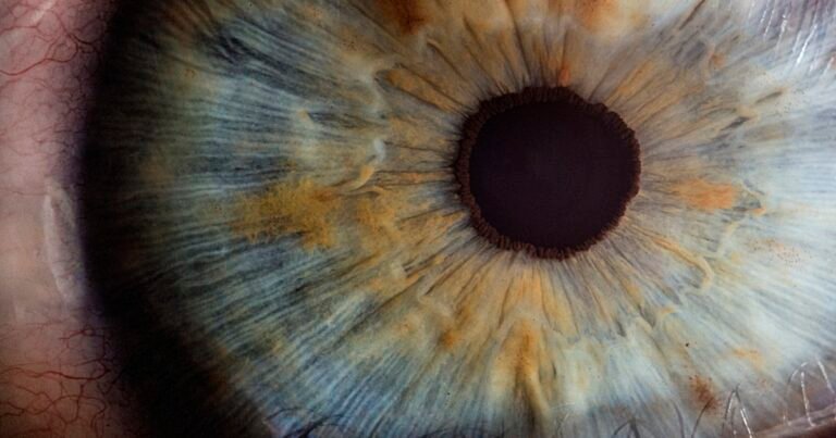
Keratoconus is a disease of the cornea that most often appears during adolescence or in young adults. It progressively deforms the cornea, which thins and takes on a cone shape, impairing vision. Caught early, it can be stabilised with a treatment called corneal cross-linking. Caught late, it sometimes leads to a corneal transplant. This article will help you identify the early signs that should prompt you to seek advice, and understand the role of topographic diagnosis at the Cachan practice.
UNDERSTANDING
What is keratoconus?
Keratoconus is a non-inflammatory corneal dystrophy characterised by progressive thinning and a cone-shaped deformation of the central or paracentral part of the cornea. This change in curvature causes irregular astigmatism, often associated with progressive myopia, which beyond a certain stage can no longer be properly corrected with glasses.
The disease usually begins between the ages of 10 and 20, progresses for 10 to 20 years, then stabilises spontaneously around the fourth decade in most cases. Recent epidemiological studies put the global prevalence at around 1 case in 750 people, with significant geographic variation (up to 1 in 250 in some Middle Eastern and South Asian populations). Its frequency is probably underestimated in older literature, because today’s screening tools reveal more early or subclinical forms.
Keratoconus affects both eyes in the vast majority of cases, but often asymmetrically: one eye may appear normal while the other already shows clear signs.
SYMPTOMS
The early signs that should raise concern
Distorted vision despite a recent correction
The first sign is often visual discomfort that persists despite new glasses. Patients describe distorted outlines, ghost images (a streak instead of a point of light), or a feeling that the correction does not “hold” for long. The cause is the irregular astigmatism induced by the corneal deformation: no pair of glasses can fully correct it.
Halos and light sensitivity
Many young affected patients report halos around headlights at night, increased sensitivity to bright light and difficulty driving in the evening. These symptoms reflect the higher-order optical aberrations (coma) generated by the irregular cornea.
Frequent changes in glasses prescription
A refraction that changes every 6 to 12 months, with an increase in astigmatism and myopia in a teenager or young adult, is a red flag. If your optician adjusts the same pair twice in a year to update the values, mention it to an ophthalmologist.
Progressive astigmatism after age 16
Physiological astigmatism normally stabilises after adolescence. A progression of astigmatism after age 16-18, especially if it is asymmetric between the two eyes, should suggest early keratoconus and lead to a corneal topography.
Other clues may be added: chronic eye itching, a history of allergy or eczema, a family history of keratoconus, and above all a habit of rubbing the eyes.
The key role of eye rubbing
Repeated eye rubbing is now considered one of the main environmental risk factors for keratoconus. The mechanical stresses placed on the cornea weaken its collagen architecture and promote both the onset of the disease and its worsening in predisposed individuals.
Several clinical studies have shown a strong correlation between the intensity of rubbing and the severity of keratoconus, and some patients stop progressing after simply stopping rubbing their eyes. This measure, free and accessible, is an integral part of management.
The usual causes of rubbing in young adults are:
- eye allergies (seasonal allergic conjunctivitis, rhinitis),
- dry eye (screens, contact lenses),
- tics or habits established since childhood,
- atopic eczema of the eyelids.
Treating the cause (anti-allergy eye drops, artificial tears, eyelid hygiene) is just as important as the corneal treatment itself.
DIAGNOSIS
Diagnosis: corneal topography
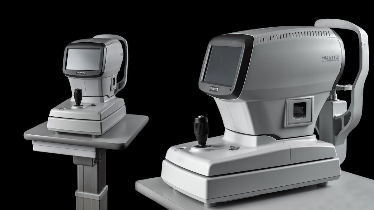
The standard eye examination (acuity, slit lamp, fundus) is not enough to diagnose early keratoconus. The diagnosis relies on high-resolution corneal imaging.
Corneal topography by Scheimpflug tomography (such as the Pentacam) is the reference standard. It analyses:
- the curvature of the anterior and posterior surfaces of the cornea,
- corneal thickness (pachymetry) point by point,
- the elevation of both surfaces relative to a reference sphere.
Several composite indices guide the diagnosis, in particular the Belin/Ambrosio Enhanced index (BAD-D), which combines posterior elevation, thickness asymmetry and pachymetric progression. A value above a defined threshold (often 1.6) points towards a suspicion of keratoconus, even when the cornea appears clinically normal.
Corneal OCT completes this analysis by measuring epithelial thickness. The epithelium tends to compensate for the stromal bulging by thinning at the apex of the cone: an asymmetric epithelial map can reveal subclinical keratoconus (forme fruste) even before it becomes visible on conventional topography.
At the Cachan practice, I have the tools needed to make this diagnosis and, above all, to track its progression over time, which is essential: it is the progression over 6 to 12 months that determines the need for treatment.
Who to see and when?
A specialist consultation is recommended if you have any of the following:
- you are under 30 and your glasses prescription is changing rapidly,
- your astigmatism increases from one visit to the next,
- a close family member has keratoconus,
- you rub your eyes regularly and have chronic allergies,
- you are considering refractive surgery and preoperative screening is essential.
Corneal topography is an integral part of the preoperative assessment I carry out before any discussion of refractive surgery: it allows a major contraindication to be ruled out before considering LASIK or PRK.
TREATMENT
Treatments according to the stage
Management depends on the stage of the disease, how fast it is progressing and the patient’s age.
Rigid or hybrid lenses (visual rehabilitation)
When the irregular astigmatism can no longer be compensated by glasses, rigid gas-permeable, scleral or hybrid lenses make it possible to restore clear vision. They do not cure the disease: they compensate for the deformation by creating a new, regular optical surface above the cornea.
Corneal cross-linking (CXL): stabilising the progression
Cross-linking is the reference treatment for stabilising progressive keratoconus. The principle: apply riboflavin (vitamin B2) to the cornea, then expose it to UV-A light to create additional bonds between the collagen fibres, which stiffens the tissue.
Randomised clinical trials and meta-analyses published in recent years show that cross-linking slows or stops progression in the majority of cases treated at the progressive stage, with an acceptable tolerability profile. The procedure is performed on an outpatient basis under topical anaesthesia, and visual recovery takes place over a few weeks.
The indication rests on documenting progression (a change in maximum keratometry, pachymetry or topographic indices) across two examinations spaced apart.
Intracorneal rings
Intracorneal rings (ring segments inserted into the corneal stroma) modify the geometry of the cornea and can improve corrected vision in some patients at a moderate stage, with or without cross-linking. They do not stop the progression of the disease.
Corneal transplant (advanced forms)
When the cornea becomes too thin, too deformed or opacified (scarred hydrops), a corneal transplant — deep anterior lamellar keratoplasty (DALK) or penetrating keratoplasty — remains the last-resort option. Thanks to early screening and cross-linking, it now concerns only a minority of patients.
In my care pathway, corneal surgery is performed at the Clinique Laser Victor Hugo, a technical facility suited to this type of procedure.
TREATMENT
Keratoconus and refractive surgery
A crucial point: keratoconus is an absolute contraindication to LASIK. Cutting the corneal flap and photoablating the stroma weaken the cornea and accelerate the deformation, with a potentially major risk of post-LASIK ectasia. No exception is made, even for a keratoconus that appears stable.
For this reason, every candidate for refractive surgery undergoes systematic topographic screening: see our dedicated page LASIK: who is it for? which details the eligibility criteria.
PRK (photorefractive keratectomy) may be discussed in certain very specific situations: forme fruste or very mild keratoconus, stable for several years, after cross-linking, and with sufficient pachymetry. This indication remains rare and requires a thorough case-by-case evaluation.
For patients with established keratoconus who wish to do without glasses, other options may be considered (scleral lenses, rings, phakic implants in some cases). The discussion always takes place after the disease has stabilised.
Living with keratoconus day to day
Having keratoconus does not condemn you to blurred vision for life. A few common-sense principles help limit progression and preserve vision:
- Stop rubbing your eyes completely, even occasionally. This is the most important measure.
- Treat eye allergies actively: antihistamine eye drops, avoidance of allergens, nasal hygiene.
- Fight dryness (artificial tears, breaks from screens, hydration).
- Have regular follow-up of your corneal topography, generally every 6 to 12 months during the progressive phase.
- Have your lenses fitted by a contact lens specialist trained in keratoconus.
The vast majority of patients diagnosed young and well monitored retain functional vision for life. The key is timing: the earlier the diagnosis is made, the more effective stabilisation is.
FAQ
Frequently asked questions
Is keratoconus hereditary?
There is a genetic component: having a first-degree relative affected increases the risk (estimated at between 10 and 20% depending on the series). This is why screening is advised for the children of affected patients, especially from adolescence onwards. The disease is not strictly hereditary, however: environmental factors (rubbing, allergy) play a decisive role.
Can you wear contact lenses with keratoconus?
Yes, and it is often essential to restore clear vision. Rigid gas-permeable, hybrid or scleral lenses are suited to deformed corneas. Fitting them requires patience and an experienced contact lens specialist, but the visual result is generally very satisfactory.
Is LASIK possible if my keratoconus is stabilised?
No. LASIK is strictly contraindicated in cases of keratoconus, even stabilised, because of the risk of postoperative ectasia. PRK on a forme fruste keratoconus that is stable and has had cross-linking may be discussed in very selected cases, but remains an exceptional indication that requires a thorough evaluation.
At what age does keratoconus appear?
Keratoconus typically begins between the ages of 10 and 25. Its detection can be later, especially for slowly progressing forms. Progression is generally most marked between 15 and 25 years of age, then tends to slow down around 30-40 years.
Is cross-linking painful?
The procedure itself is painless (anaesthesia with eye drops). The first few days afterwards can be uncomfortable, comparable to a corneal abrasion (a sensation of grit, watering, light sensitivity), especially for the “epi-off” technique that removes the epithelium. A bandage contact lens and a pain-relief treatment are prescribed for this period.
Can my keratoconus stop progressing on its own?
Yes. Many cases of keratoconus stabilise naturally around 30-40 years of age. It is precisely the documentation of stability (or, conversely, of progression) on topography that guides the decision whether or not to offer cross-linking. Not all keratoconus needs to be treated: some remain stable and compensated by glasses or lenses for a lifetime.
Scientific sources
- Santodomingo-Rubido J, Carracedo G, Suzaki A, Villa-Collar C, Vincent SJ, Wolffsohn JS. Keratoconus: An updated review. Contact Lens and Anterior Eye. 2022;45(3):101559. PMID 34991971.
- McMonnies CW. Mechanisms of rubbing-related corneal trauma in keratoconus. Cornea. 2009;28(6):607-15. PMID 19512912.
- Wittig-Silva C, Chan E, Islam FM, Wu T, Whiting M, Snibson GR. A randomized, controlled trial of corneal collagen cross-linking in progressive keratoconus: three-year results. Ophthalmology. 2014;121(4):812-21. PMID 24393351.
- Bamdad S, Sedaghat MR, Yasemi M, Vahedi A. Sensitivity and Specificity of Belin Ambrosio Enhanced Ectasia Display in Early Diagnosis of Keratoconus. Journal of Ophthalmology. 2020. PMID 33489342.
- Gomes JAP, Tan D, Rapuano CJ, et al. Group of Panelists for the Global Delphi Panel of Keratoconus and Ectatic Diseases. Global consensus on keratoconus and ectatic diseases. Cornea. 2015;34(4):359-69. PMID 25738235.
Further reading
- Keratoconus: diagnosis and treatment — complete management according to the progressive stage
- LASIK: who is it for? — why LASIK is contraindicated in cases of keratoconus
- Preoperative assessment — corneal topography during the consultation
- Book an appointment — specialist consultation in Cachan
Cachan practice · Tel. 01 45 47 08 11
Disclaimer
This article is for information purposes. A personalised ophthalmological opinion remains essential for any treatment decision.
This content is written for information purposes and is not a substitute for a medical consultation. Diagnosing keratoconus and deciding on a treatment (cross-linking, rings, transplant) rests on a clinical examination and corneal imaging carried out by an ophthalmologist. The progression timelines, treatment choices and eligibility factors described in this article are generic and must be adapted to your individual situation. In the event of a rapid drop in vision, pain or new discomfort, seek advice without delay.
For a personalised evaluation, you can book an appointment at the Cachan practice via Doctolib: https://www.doctolib.fr/ophtalmologue/cachan/moise-tourabaly.
Written and reviewed by Dr Moïse Tourabaly, ophthalmic refractive surgeon — former chief resident (Quinze-Vingts National Eye Hospital).
Last updated: July 6, 2026