Light halos around lights: causes, emergencies and when to seek care
A light halo is a ring or aura perceived around a light source — car headlights, street lamps, screens — especially at night. This phenomenon is often harmless, linked to dry eye or contact lens wear, but it can also signal an early cataract, keratoconus or, in an acute picture with pain and a red eye, an attack of angle-closure glaucoma: an ophthalmic emergency. Understanding the origin of halos makes it possible to know whether you should seek urgent care or attend a routine appointment.
UNDERSTANDING
What is a light halo?
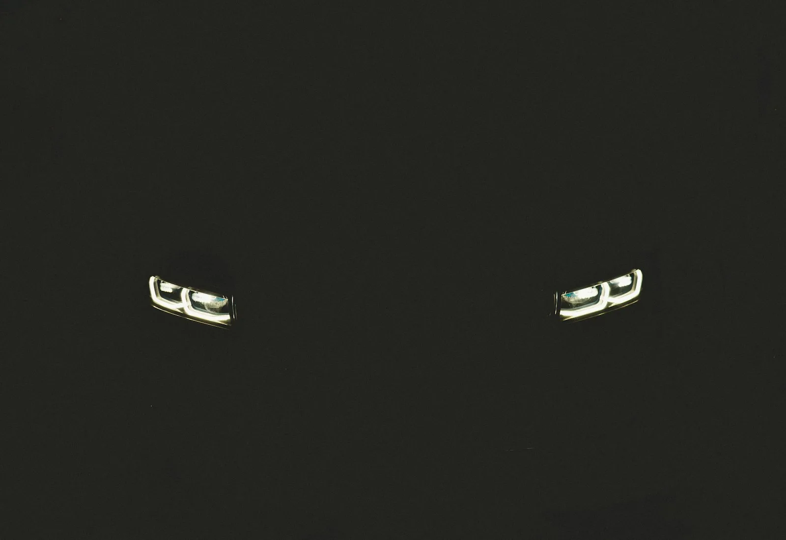
A light halo is a diffuse circle, an aura or a corona perceived around a point of light. It typically appears in the evening or in darkness, when the pupil dilates to gather more light: car headlights, traffic lights, street lamps or screens are then surrounded by a blurred, sometimes iridescent ring.
The mechanism is optical. Normally, light passes through the cornea and then the crystalline lens to be focused to a sharp point on the retina. When one of these transparent media becomes slightly irregular or cloudy — an unstable corneal surface, an opacified lens, a post-surgical scar — the light is scattered instead of being focused, creating the characteristic ring.
Pupil size plays a key role: the wider it is, the more peripheral rays (where optical imperfections are most pronounced) enter the eye. This is why halos are almost always more noticeable at night than during the day.
Halos, phosphenes, floaters: three distinct symptoms
These three phenomena are often confused, but their causes differ:
- Light halo: a ring or aura surrounding a real light source. Corneal, lenticular or pupillary in origin.
- Phosphenes: flashes or bursts of light, often perceived to the side, without an external light source. Linked to vitreoretinal traction or mechanical stimulation of the retina.
- Floaters: filaments, dots or cobweb shapes that follow the movement of your gaze. Vitreous in origin in almost all cases.
BENIGN CAUSES
Benign and common causes of light halos
Most nocturnal halos have a simple and reversible explanation. Here they are in order of frequency:
Dry eye
The tear film forms the first optical surface of the eye. When it is unstable or insufficient — functional dry eye, prolonged screen exposure, air conditioning, contact lens wear — the corneal surface becomes irregular and scatters light. Halos linked to dry eye are characteristic: they fluctuate within seconds and ease markedly after a blink or the instillation of artificial tears.
Large pupil at night
In darkness, the pupil dilates to gather more light. It then lets rays enter through the periphery of the cornea and lens, where the natural optical aberrations are greater. People who naturally have large pupils in scotopic conditions perceive more halos at night, without this reflecting any disease. This is also why measuring the pupil diameter forms part of the pre-operative assessment before any refractive surgery.
Contact lens wear
Poorly fitted, soiled or dried-out lenses at the end of the day blur the optical surface and generate halos. A decentred lens or one with too small an optical zone accentuates the phenomenon. A fitting check by the ophthalmologist usually makes it possible to correct the discomfort by changing the type or material of the lens.
Uncorrected hyperopia and astigmatism
An under-corrected or uncorrected refractive error — especially in low-light conditions where pupillary dilation makes focusing more difficult — may manifest as diffuse halos around light sources. A refraction check and the fitting of an appropriate optical correction are sufficient in this case.
CAUSES TO INVESTIGATE
Causes that warrant an ophthalmology consultation
Early cataract
The progressive opacification of the crystalline lens — the cataract — scatters light instead of focusing it, causing halos, marked glare when facing headlights and a sense of fog, especially at night. These cataract-related halos develop slowly, over months or years, and are accompanied by other signs: reduced distance visual acuity, an increased need for light to read, colours that appear dull or yellowed. If you recognise this picture, the page on the symptoms of cataract details the warning signs.
Keratoconus
Keratoconus is a progressive deformation of the cornea, which takes on a cone shape rather than a spherical one. This irregularity of the corneal surface generates halos, double vision (monocular diplopia) and fluctuating blur, especially at night. It preferentially affects young adults. Diagnosis relies on corneal topography.
Corneal oedema
A swelling of the layers of the cornea — corneal oedema — disrupts its transparency and creates significant halos, often more pronounced in the morning (the cornea dehydrates less at night). It can arise in several contexts: prolonged lens wear, corneal dystrophy, post-operative complication, or ocular hypertension.
Chronic ocular hypertension and glaucoma
Chronic open-angle glaucoma may be accompanied by halos, particularly when the intraocular pressure is high and causes slight corneal oedema. These chronic, undramatic halos form part of a broader picture: raised ocular pressure, optic disc cupping, visual field damage. Distinguishing this from an acute attack (see box below) is essential.
Emergency: acute angle-closure glaucoma attack
The combination of sudden light halos + intense ocular pain + red eye + reduced vision + nausea or vomiting constitutes an absolute ophthalmic emergency. This picture suggests an attack of acute angle-closure glaucoma: the intraocular pressure rises abruptly (sometimes above 50 mmHg), compressing the optic nerve.
What to do: call the emergency services or go immediately to an ophthalmic emergency department. Every hour counts to preserve the optic nerve. Do not wait until the following day.
REFRACTIVE SURGERY
Halos after refractive surgery: is this normal?
Yes, and it is an expected and documented situation. After LASIK or PKR, halos and sensitivity to glare are common during the first few weeks. They are explained by the healing of the cornea and by the transition between the treated zone and the untreated peripheral cornea, more visible when the pupil dilates at night.
In the great majority of cases, these halos diminish gradually over several weeks to several months, with corneal healing and visual neuroadaptation. They do not always fade completely, but their intensity eases markedly. Transient post-operative dry eye contributes to them and is treated with frequent artificial tears.
A careful pre-operative assessment reduces this risk: measurement of the pupil diameter in low light, analysis of corneal topography, centring of the treatment zone. Halos that persist or worsen several months after the procedure warrant a check to find the precise cause — oedema, refractive regression, persistent dryness.
Key points on halos after LASIK
Nocturnal halos after refractive surgery are transient in the great majority of cases. Measuring the pupil in scotopic conditions before the operation makes it possible to personalise the treatment zone and reduce their intensity. If you are considering refractive surgery, the pre-operative assessment systematically includes this measurement.
EXAMINATION
How are light halos investigated?
Faced with halos, the ophthalmologist first seeks to clarify the clinical context: for how long, in one or both eyes, with or without pain, associated with other visual signs. This history-taking already points towards the diagnosis. The examination then continues through several complementary steps:
- Measurement of visual acuity and refraction, to detect reduced vision or an under-corrected error.
- Slit-lamp examination: analysis of the cornea (surface, transparency, regularity), the tear film and the lens (early cataract).
- Measurement of intraocular pressure (tonometry), essential when halos are associated with pain or a red eye.
- Study of the tear film (Schirmer test, tear break-up time) to look for dryness.
- Fundus examination after dilation, if glaucoma or a retinal condition is suspected.
- Corneal topography, particularly to detect keratoconus or to analyse an operated cornea.
Depending on the context, a measurement of the pupil diameter in scotopic conditions, corneal pachymetry or an examination of the iridocorneal angles (gonioscopy) may complete the assessment. The aim is to identify the precise cause in order to propose appropriate management: treatment of dryness, optical fitting, or referral for surgery (cataract, treatment of keratoconus).
WHEN TO SEEK CARE

When to seek care and how urgently?
The answer depends above all on the associated symptoms:
Immediate emergency (call the emergency services or go to A&E)
- Halos that appear suddenly in one eye, within a few hours
- Intense ocular pain, radiating to the forehead or temple
- A red, hard eye, with blurred or severely reduced vision
- Associated nausea or vomiting
- Halos associated with a shower of moving black dots or with recent, sudden flashes of light (phosphenes)
Scheduled consultation within 2 to 4 weeks
- Stable, bilateral nocturnal halos, without pain, that appeared gradually
- Fluctuating halos, eased by blinking or artificial tears (dryness likely)
- Halos that appeared after starting contact lens wear
- Post-operative halos (LASIK, PKR) present for more than 3 months without improvement
- Halos associated with a progressive decline in night vision (possible early cataract)
FAQ
Frequently asked questions about light halos
Are light halos always serious?
No. Most of the time, halos are harmless: dry eye, a large pupil at night, tired lenses or uncorrected hyperopia. They become concerning when they appear suddenly in one eye and are accompanied by pain, a red eye or a rapid decline in vision. In this latter case, seek urgent care.
Why do I see halos around headlights at night?
At night, the pupil dilates to gather more light. It then lets rays enter through the periphery of the eye, where the natural optical imperfections are greater. Dry eye, a large pupil or an early cataract accentuate this phenomenon. An ophthalmology examination makes it possible to identify the cause and propose appropriate management.
Do halos after LASIK or PKR go away?
Nocturnal halos are common in the first few weeks after LASIK or PKR. They diminish gradually over several weeks to several months, with corneal healing and neuroadaptation. They do not always fade completely, but their intensity eases in the majority of patients. Halos that persist or worsen beyond 3 months should be assessed.
Halo, phosphene or floater: what is the difference?
A halo is a ring perceived around a real light source. A phosphene is a flash or burst of light, often lateral, without an external source. A floater is a filament or mobile dot that follows your gaze. These symptoms have different mechanisms: an examination makes it possible to distinguish them and to look for the cause.
Can halos come from glaucoma?
Yes, in two quite different contexts. Chronic open-angle glaucoma can cause faint halos linked to raised pressure and slight corneal oedema. An acute glaucoma attack, by contrast, causes sudden and intense halos, associated with severe pain, a red and hard eye, and a rapid decline in vision: this is an absolute emergency that requires contacting the emergency services or going immediately to A&E.
Dry eye and halos: what is the link?
Dry eye is one of the most common causes of nocturnal halos. An unstable tear film makes the corneal surface irregular and scatters light. The characteristic sign: the halos fluctuate and ease immediately after a blink or the instillation of artificial tears. Treating the dryness is usually enough to make them regress.
Can a light halo be caused by keratoconus?
Yes. Keratoconus deforms the cornea and creates surface irregularities that scatter light in multiple directions, causing halos, double vision (monocular diplopia) and fluctuating blur — often more pronounced at night. Diagnosis relies on corneal topography. If you are under 35 and perceive halos that are gradually increasing, a consultation is required to rule out this diagnosis.
Sources
- French Society of Ophthalmology (SFO) — Semiology of visual disturbances: halos, glare, cataract, refractive surgery and ophthalmic emergencies.
- College of University Ophthalmologists of France (COUF) — Crystalline lens and cataract, ocular surface and tear film, glaucoma, ophthalmic emergencies.
- French National Authority for Health (HAS) — Recommendations on corneal refractive surgery and on the management of acute angle-closure glaucoma.
- Ameli.fr (French Health Insurance) — Patient factsheets: dry eye, warning signs in ophthalmology, what to do when facing an ocular emergency.
This article is intended for information purposes and does not replace a medical consultation. Only an ophthalmology examination can determine the cause of your light halos, establish a personalised diagnosis and direct you towards appropriate management.
Are halos bothering or worrying you?
Dr Moïse Tourabaly, former chef de clinique at the Quinze-Vingts hospital, carries out a complete assessment of your vision, measures the ocular pressure, and analyses the cornea and the lens to identify the cause of your light halos and propose appropriate management.
Written and reviewed by Dr Moïse Tourabaly, ophthalmic refractive surgeon — former chief resident (Quinze-Vingts National Eye Hospital).
Last updated: July 27, 2026