Visual acuity: definition, measurement and interpretation (20/20)
Visual acuity is the eye’s ability to distinguish fine detail and to separate two very close points. In France it is expressed in tenths on the Monoyer chart, where 10/10 is the reference for normal vision; the equivalent in English-speaking countries is 20/20 on the Snellen chart. Understanding what this figure means, what it measures, what it does not tell you, and how it relates to optical errors helps you interpret your eye assessment and talk things through with your ophthalmologist.
UNDERSTANDING
What is visual acuity?
Visual acuity refers to the resolving power of the eye: its ability to perceive the smallest details of an object and to distinguish two points separated by a very small angular gap. In practice, it is what allows you to read the letters on a road sign, recognise a face from a distance, or make out small print.
Two complementary types of acuity are distinguished: distance acuity, measured at several metres, which reflects vision at a distance; and near acuity, assessed at reading distance (around 33 cm). Both can be impaired independently. Good distance acuity does not rule out difficulty with near vision, particularly after the age of 45 with the onset of presbyopia.
Acuity depends on several factors: the transparency of the optical media (cornea, lens), the precision of focus of the image on the retina, and the integrity of the retina and visual pathways up to the visual cortex. A drop in acuity may therefore stem from a simple correctable optical error or from an eye disease requiring treatment.
MEASUREMENT
How is visual acuity measured?
Visual acuity is measured using standardised charts of letters, numbers or symbols of decreasing size, presented at a set distance. The patient reads the smallest line they can make out, one eye at a time, first without correction and then with. The result reflects the level of detail the eye is able to resolve.
The Monoyer chart (distance vision, France)
In France, distance vision is assessed with the Monoyer chart, read at 5 metres. It has lines of letters graded from 1/10 to 10/10. Reading the 10/10 line corresponds to acuity considered normal. This notation in tenths is the everyday language of French ophthalmologists and appears in every eye-assessment report.
The Snellen chart (20/20, English-speaking countries)
In English-speaking countries, the Snellen chart is used, measured at 20 feet (about 6 metres). Normal vision is recorded there as 20/20, which is exactly equivalent to the French 10/10. A result of 20/40 means the patient sees at 20 feet what a normal eye sees at 40 feet: an acuity of 5/10. Both systems describe the same reality with different units.
The Parinaud chart (near vision)
For near vision, the Parinaud chart is used, a set of paragraphs in decreasing type sizes read at about 33 cm. The result ranges from Parinaud 14 (large type) to Parinaud 2 (small type), with Parinaud 2 corresponding to good reading vision. This measurement is essential for assessing presbyopia and guiding the prescription of a reading addition.
Refraction, an indispensable preliminary
The acuity measurement is accompanied by a refraction: the ophthalmologist determines the optical correction, expressed in dioptres, that gives the highest possible acuity. A distinction is made between uncorrected acuity (spontaneous acuity) and corrected acuity. A short-sighted eye may have only 2/10 without glasses and reach 10/10 once corrected, which points to a pure refractive error, with no associated disease.
Key point
Acuity is always measured with and without correction. If acuity returns to 10/10 with the right optical correction, the cause of the blur is refractive. If it stays low despite correction, a disease of the retina, the optic nerve or the optical media must be looked for.
INTERPRETATION
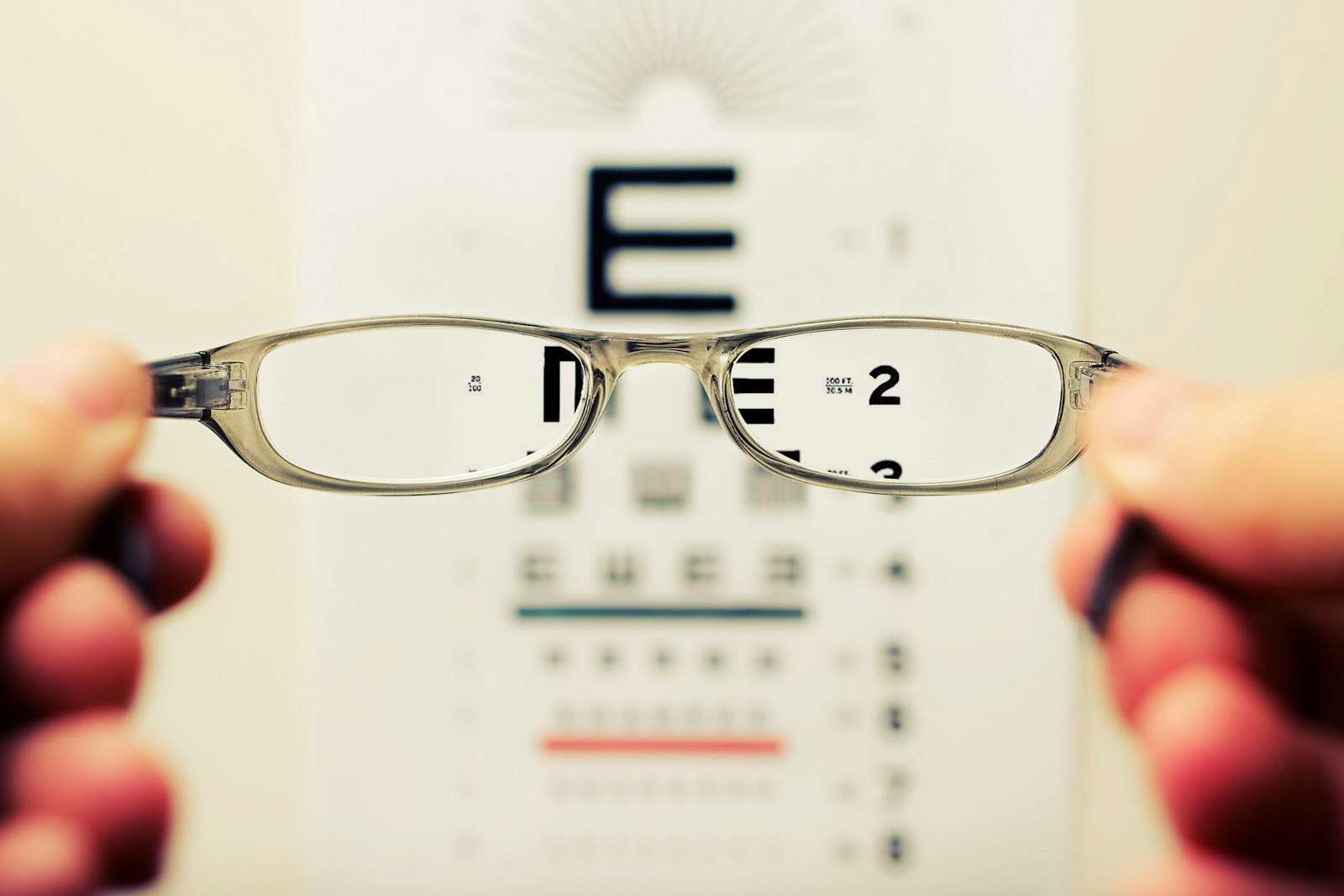
What does 20/20 mean? What this figure says, and what it does not
10/10 corresponds to normal visual acuity: the person reads the smallest standard line of the Monoyer chart at 5 metres. Its English-speaking equivalent is 20/20 on the Snellen chart. These two notations denote exactly the same level of performance and serve as the statistical reference for normal vision in the population.
One misconception needs clearing up, however: 10/10 does not mean perfect, absolute vision. It is a statistical threshold of normality, not a theoretical maximum. Some people read beyond it, 12/10 or more, without this reflecting extraordinary vision. Conversely, acuity of 10/10 does not rule out other visual problems: reduced contrast sensitivity, poor night vision, a peripheral visual-field defect or sensitivity to glare.
Visual acuity is therefore only one of the parameters of visual quality. A complete eye assessment also evaluates the visual field, colour vision, contrast sensitivity and the health of the retina. The acuity figure nonetheless remains the simplest and most universal indicator for an initial clinical benchmark.
ACUITY AND REFRACTION
Visual acuity and optical errors: myopia, hyperopia, astigmatism, presbyopia
Optical errors, or refractive errors, are the most common causes of low visual acuity. Each has a characteristic pattern of difficulty. Understanding how they relate to acuity helps you interpret the figures in your assessment and anticipate the benefit expected from a correction.
Myopia: reduced distance acuity
In the short-sighted patient, the image of a distant object forms in front of the retina. Uncorrected distance acuity is therefore reduced, sometimes severely, while near acuity often stays normal or is little affected. A myope of −3.00 dioptres may have only 2 to 3/10 in the distance without glasses, then return to 10/10 with correction. The stronger the myopia, the greater the drop in uncorrected acuity.
Hyperopia: eye strain and variable blur
Hyperopia (long-sightedness) is more deceptive: the eyeball is too short and the image forms behind the retina. Young adults often compensate through accommodation (contraction of the lens), which can mask the error for years, with acuity of 10/10 despite the refractive problem. But this constant compensation causes eye strain and headaches. With age, as accommodation declines, near acuity deteriorates first.
Astigmatism: blur at all distances
Astigmatism results from a cornea whose curvature is not perfectly spherical. Unlike myopia or hyperopia, it impairs acuity both at distance and at near: images appear distorted or doubled along a particular axis. Moderate astigmatism (−1.50 dioptres) can noticeably lower visual quality and explain difficulty reading small print even when the sphere is well corrected.
Presbyopia: near acuity that declines after 45
Presbyopia is not a defect of the eye in the strict sense, but an ageing of the lens, which gradually loses its elasticity. Accommodation decreases, and near acuity deteriorates from about the age of 45. Distance acuity, on the other hand, is not affected by presbyopia alone. The typical presbyopic patient sees well in the distance but has to hold text further and further away to read it.
ACUITY VS OTHER PARAMETERS
Visual acuity, visual field, refraction: three complementary measurements
Visual acuity is often confused with other parameters that describe vision from different angles. These three measurements are complementary and cannot be substituted for one another.
Visual acuity measures central sharpness
Visual acuity measures only central vision: the ability to see fine detail in the line of sight. It says nothing about peripheral vision, nor about the ability to detect movements or objects to the side. Normal acuity of 10/10 is perfectly compatible with severe visual-field damage.
The visual field measures the extent of vision
The visual field assesses the area of the visible world without moving the eyes: a normal eye perceives information over about 180° horizontally. Exploring it is essential for detecting glaucoma (which erodes the periphery first) and other conditions of the optic nerve. A patient may keep acuity of 10/10 while having a very restricted visual field, the “tunnel vision” of advanced glaucoma.
Refraction measures the correction required
Refraction quantifies the power of optical correction, in dioptres, that the eye needs to form a sharp image on the retina. It is the measurement that appears on a glasses prescription. It is not directly linked to acuity: two people with the same correction in dioptres can have very different acuities depending on the state of their retina or the presence of amblyopia.
In brief
Acuity (10/10), visual field and refraction (dioptres) are three distinct tools. Acuity measures central sharpness; the visual field measures peripheral extent; refraction measures the power of correction required. A complete assessment explores all three.
WHEN TO SEE A DOCTOR
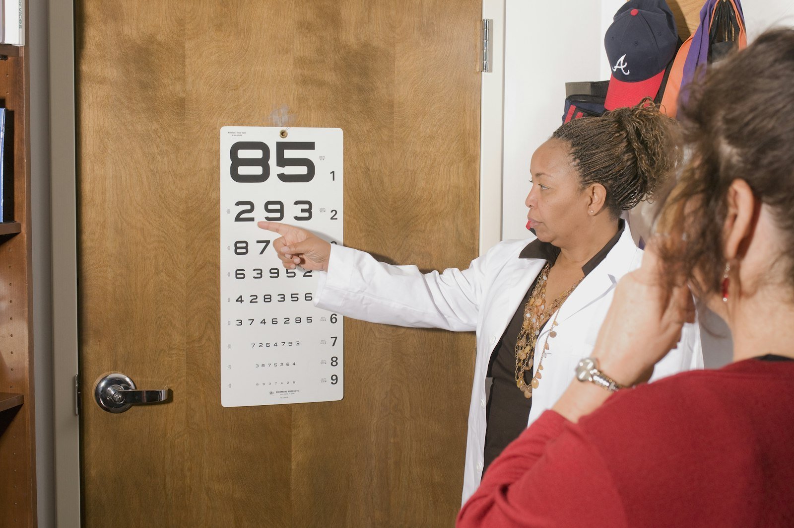
When should you see a doctor about a drop in visual acuity?
A drop in visual acuity falls into two broad families of causes: a refractive error, which is corrected with lenses, or an eye disease, which requires specific treatment. The distinction is made simply during an examination: if acuity returns to 10/10 with the right correction, the cause is refractive; if not, a disease must be looked for.
- Gradual decline in distance vision: often myopia that is beginning or worsening. A consultation is useful to adjust the correction.
- Eye strain and headaches at the end of the day: suggestive of poorly corrected hyperopia or astigmatism, particularly in children.
- Growing difficulty reading close up after 45: a sign of presbyopia. An assessment can establish the appropriate correction.
- Rapid or sudden loss in one or both eyes: an ophthalmic emergency. It may signal a retinal detachment, a vascular occlusion, optic neuritis or another serious condition.
- A veil, distortion or spot in the central visual field: may suggest macular involvement (early AMD), see a doctor without delay.
For stable refractive errors, surgery may be an option depending on the patient’s profile. Feasibility is determined during a complete pre-operative assessment including corneal topography, pachymetry and measurement of the anterior chamber. The choice between LASIK, PRK, SMILE or a phakic implant depends on the corneal profile and the error to be corrected.
FREQUENTLY ASKED QUESTIONS
Frequently asked questions about visual acuity
Does 20/20 mean I have perfect vision?
No. 10/10 (20/20) is a statistical threshold of normality, not an absolute maximum. Some people read beyond it, 12/10 or more, on the measurement charts. Moreover, acuity of 10/10 does not rule out other visual problems: reduced contrast sensitivity, a peripheral visual-field defect or difficulty with glare. A complete assessment always explores several complementary parameters.
What visual acuity do you need to drive in France?
French regulations (the order on fitness to drive) require binocular acuity of at least 5/10, with correction if necessary, for a standard category B licence. Specific conditions apply in cases of monocular vision. Only a formal medical evaluation can confirm fitness to drive; the figures from an in-practice assessment do not amount to an administrative certificate.
Visual acuity and dioptres: how are they related?
They are two independent measurements. Acuity (in tenths) measures actual visual performance, what the eye manages to distinguish. The dioptre measures the power of optical correction needed to compensate for a refractive error. You can have −6.00 dioptres of myopia and see 10/10 with your glasses, or have only −1.00 dioptre yet see only 7/10 because of amblyopia. The link is not a direct one.
Can you have better than 20/20?
Yes. 10/10 (20/20) is a threshold of normality, not an absolute physiological limit. Some people, particularly the young and those without any optical error, reach 12/10 or more on the measurement charts. This simply reflects a resolving ability slightly above the average of the reference population used to establish the chart.
How is a child’s visual acuity measured?
In young children who cannot yet read, adapted charts are used: pictures, symbols or letters to match (the tumbling-E test, images to name). In infants, the ophthalmologist relies on behavioural tests and Teller acuity cards. Early screening is essential to spot amblyopia (lazy eye) before the age of 6 to 7, the cut-off period for effective treatment.
Does visual acuity decline with age?
Near vision inevitably changes with presbyopia, from about the age of 45, because of ageing of the lens. Distance vision, on the other hand, does not necessarily decline: well corrected, it can stay normal for a long time. A drop in distance acuity in an adult is not an inevitable consequence of age and should prompt a search for a cause, early cataract, retinal disease or something else.
Does good visual acuity mean I do not have glaucoma?
No, and that is precisely the danger of glaucoma: it can gradually destroy the peripheral visual field over years without affecting central acuity, which stays at 10/10. At an advanced stage, vision becomes tubular (only the centre is preserved) before acuity itself falls. Screening for glaucoma relies on measuring eye pressure, examining the optic nerve and exploring the visual field, not on acuity alone.
Sources
- French Society of Ophthalmology (SFO) — Measurement of visual acuity: principles of the Monoyer, Snellen and Parinaud charts.
- College of University Ophthalmologists of France (COUF) — Item “Alteration of visual function”: acuity, refraction, visual field and causes of a drop in acuity.
- French National Authority for Health (HAS) — Screening and management of visual disorders in children and adults.
- Ministry of the Interior / Road Safety — Order on the visual conditions required for the issue of a driving licence.
This article is for information only and does not replace a medical consultation. Only a complete ophthalmic examination can measure your visual acuity, interpret the result in its context and establish a personalised diagnosis.
Has your visual acuity dropped?
Dr Moïse Tourabaly, former chief resident at the Quinze-Vingts hospital, measures your acuity, looks for the cause and advises you on the solutions best suited to your situation, whether optical correction or refractive surgery.
Written and reviewed by Dr Moïse Tourabaly, ophthalmic refractive surgeon — former chief resident (Quinze-Vingts National Eye Hospital).
Last updated: July 8, 2026