HbA1c and eye health: the link between blood sugar and vision
Glycated hemoglobin — HbA1c — is the reference marker for blood sugar control over the past three months. Beyond being a figure for endocrinology follow-up, it is also a major determinant of visual prognosis in people with diabetes. Here is what this lab result tells you about your retina, and how ophthalmological monitoring fits in with this benchmark.
UNDERSTANDING
Understanding HbA1c
HbA1c (glycated hemoglobin, sometimes written A1C) corresponds to the fraction of hemoglobin to which glucose has bound through a non-enzymatic reaction. Since the lifespan of a red blood cell is about 120 days, this measurement reflects the average blood sugar over the past 2 to 3 months — a more stable snapshot than a single fasting blood glucose reading.
The thresholds used in France and recognized internationally are as follows:
- HbA1c < 5.7%: normal
- HbA1c between 5.7% and 6.4%: prediabetes
- HbA1c ≥ 6.5% (on two readings): diabetes
The result is reported as a percentage and sometimes, in addition, in mmol/mol according to IFCC standardization. This measurement is performed with a simple blood draw, with no need to fast.
WHO IS CONCERNED
Who is concerned?
HbA1c monitoring mainly concerns people with established diabetes, whatever its type:
- Type 1 diabetes (about 10% of diabetes cases) — an absolute insulin deficiency of autoimmune origin, requiring insulin therapy from the outset.
- Type 2 diabetes (about 90%) — insulin resistance followed by a relative insulin deficit, linked to several factors (family history, sedentary lifestyle, excess weight, age).
- Gestational diabetes — appearing during pregnancy, with an increased risk of later type 2 diabetes for the mother.
In France, about 4.2 million people were being treated for diabetes in 2023 according to Santé Publique France, or nearly 6% of the population. A significant proportion of cases probably remains undiagnosed. Whatever your type of diabetes, regular ophthalmological screening is recommended for you — in coordination with your care team.
COMPLICATIONS
Eye complications of diabetes
Diabetes exposes you to several ophthalmological complications, whose occurrence and severity are influenced by the average HbA1c level over the years. The main ones are diabetic retinopathy, diabetic macular edema, and early cataract.
Diabetic retinopathy (NPDR / PDR)
Diabetic retinopathy is damage to the capillaries of the retina. It progresses through two main stages:
- Non-proliferative retinopathy (NPDR) — microaneurysms, dot hemorrhages, exudates. Often asymptomatic, which is why screening is so important.
- Proliferative retinopathy (PDR) — a more advanced stage, with the appearance of fragile new vessels that can bleed into the vitreous or even cause the retina to detach. Prompt management (panretinal laser, intravitreal injections) is then essential.
After 15 to 20 years of diabetes, retinopathy affects a significant proportion of patients, in varying proportions depending on blood sugar control and other cardiovascular factors.
Diabetic macular edema (DME)
Key point
Diabetic macular edema (DME) is a thickening of the macula caused by leakage from the capillaries. It is today one of the leading causes of vision loss in patients with diabetes. Early diagnosis through macular OCT is decisive: modern treatment (intravitreal anti-VEGF injections, sometimes focal laser) can stabilize or even improve vision, provided it is started early.
Early diabetic cataract
Poorly controlled diabetes also promotes the appearance of a cataract earlier than in the general population. Its surgical management remains technically comparable to that of an ordinary cataract, but it requires a rigorous prior retinal assessment, particularly when an associated retinopathy is present.
TARGETS
Thresholds and treatment targets
Two landmark studies, still cited today, laid the groundwork for the link between blood sugar control and eye complications: the DCCT (type 1 diabetes) and the UKPDS (type 2 diabetes).
Key figure
Every 1% reduction in HbA1c is associated with about a 37% reduction in microvascular complications — including retinopathy (UKPDS 33, Lancet, 1998).
The DCCT (Diabetes Control and Complications Trial, NEJM, 1993) demonstrated, in people with type 1 diabetes, that intensive blood sugar control — aiming for an HbA1c around 7% — reduces the risk of developing retinopathy by about 76% and slows its progression by about 54%, compared with conventional treatment.
The UKPDS 33, published in 1998 for type 2 diabetes, established a continuous relationship: every 1% reduction in HbA1c is associated with about a 37% reduction in microvascular complications. There is no threshold below which the benefit abruptly disappears: any improvement, even a modest one, translates into clinical benefit.
In practice, the recommendations of the ADA (Standards of Care 2024) and the HAS (2013, type 2 diabetes) set a general HbA1c target below 7% in adults. This target is, however, individualized, typically between 6.5% and 8% depending on age, how long the diabetes has been present, the risk of hypoglycemia, and comorbidities. This target should be defined with your treating physician or your diabetologist.
FOLLOW-UP
Ophthalmological follow-up: OCT and OCT angiography at the Paris 13 practice
Regardless of the HbA1c figure, an annual eye examination is recommended for anyone with diabetes — more frequently if retinopathy or edema is already documented. Follow-up at the practice is organized around three stages:
- History and visual acuity — how long you have had diabetes, current treatments, latest HbA1c results, family history, recent visual symptoms (reduced acuity, floaters, distortion of lines).
- Fundus examination after pupil dilation — looking for hemorrhages, microaneurysms, exudates, new vessels.
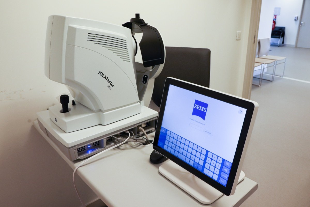
- Additional imaging — macular optical coherence tomography (OCT) to detect edema, OCT angiography to visualize retinal microcirculation without injecting any contrast agent.
I see you for these retinal assessments at my practice at 12 Rue du Moulin des Prés, Paris 13, within the Diabet’ Paris 13 team in collaboration with the area’s diabetology teams. The practice is equipped with a latest-generation swept-source OCT, well suited to monitoring the retinal conditions of diabetes, particularly as part of the screening and follow-up of diabetic retinopathy. Depending on the results, the follow-up schedule is then adjusted (annual, twice-yearly, or even more frequent).
When to seek care without delay
Certain visual signs warrant a prompt ophthalmological consultation, without waiting for the annual appointment:
- Recent vision loss, in one or both eyes
- Sudden onset of floaters or a shower of soot in the field of vision
- A dark veil or persistent shadow
- Distortion of straight lines (altered Amsler grid, wavy text)
- Associated eye pain or redness
FAQ
Frequently asked questions
Book an appointment with Dr Tourabaly
Diabetic retinal assessment at my practice at 12 Rue du Moulin des Prés, Paris 13 (Diabet’ Paris 13 team). OCT and OCT angiography available on site. Book online or by phone at 01 89 31 30 60.
Sources
- The Diabetes Control and Complications Trial Research Group. The effect of intensive treatment of diabetes on the development and progression of long-term complications in insulin-dependent diabetes mellitus. N Engl J Med. 1993;329(14):977-986. DOI : 10.1056/NEJM199309303291401 — PMID : 8366922.
- UK Prospective Diabetes Study (UKPDS) Group. Intensive blood-glucose control with sulphonylureas or insulin compared with conventional treatment and risk of complications in patients with type 2 diabetes (UKPDS 33). Lancet. 1998;352(9131):837-853. DOI : 10.1016/S0140-6736(98)07019-6 — PMID : 9742976.
- American Diabetes Association Professional Practice Committee. Retinopathy, Neuropathy, and Foot Care: Standards of Care in Diabetes — 2024. Diabetes Care. 2024;47(Suppl. 1):S231-S243. PMID 38078577.
- Haute Autorité de Santé. Stratégie médicamenteuse du contrôle glycémique du diabète de type 2. Recommandations 2013.
- Santé Publique France. Prévalence du diabète traité pharmacologiquement en France. Bulletin épidémiologique hebdomadaire, 2023.
This content is informational and does not replace a medical consultation. Each situation is individual and must be assessed with your treating physician, your diabetologist, or your ophthalmologist.
Written and reviewed by Dr Moïse Tourabaly, ophthalmic refractive surgeon — former chief resident (Quinze-Vingts National Eye Hospital).
Last updated: July 6, 2026